21 Viruses Can Be Visualized Using Which Method
Viruses are so small that they are best viewed using an electron microscope which is how they were first visualized in the 1940s. The tagged virus is harvested and infected into another cell 3.
Viruses Free Full Text Viral Infection At High Magnification 3d Electron Microscopy Methods To Analyze The Architecture Of Infected Cells Html
In plants infected with unmodified viruses the mechanism is specifically targeted against the viral genome.
. Viruses generally come in two forms. Fluorescent dyes - such as propidium iodide and. Today we know that viruses can be found nearly anywhere in the air oceans and soil.
Using VioletUV illumination the SP-IRIS technique is able to detect individual flavivirus particles 40 nm while green light illumination is capable of identifying and discriminating between vesicular stomatitis virus and vaccinia virus 360 nm. Most viruses are easily visualized with a light microscope. The complexity of packing of the RNA can be appreciated using a paper ribbon for the RNA place in the paper shell.
Combined use of TEM and SEM improves characterization of larger objects like baculovirus occlusion bodies Gencer et al 2018. Although virus crystals were investigated using both the contact and tapping modes. One method of amplification is to use a label a fluorescent dye or quantum dot that emits many photons per label instead of relying on some intrinsic optical property of the analyte molecule.
In any of these electrophoresis techniques the locations of the DNA or RNA fragments in the gel can be detected by various methods. Retrograde tracing is a research method used in neuroscience to trace neural connections from their point of termination the synapse to their source the cell bodyRetrograde tracing techniques allow for detailed assessment of neuronal connections between a target population of neurons and their inputs throughout the nervous systemThese techniques allow the mapping. Naked DNA viruses may be seen in the cytoplasm after the.
A tiny percentage of these are dangerous pathogens that cause disease such as the current coronavirus called. However with virus vectors carrying sequences derived from host genes the process can be additionally targeted. Feb 1 2014.
To visualize DiD-labeled PEDVs using an immunofluorescence assay 25 μl of viral supernatant was fixed on a glass-bottomed cell culture dish containing Polybrene 8 μgml final concentration using stock 8 mgml at 11000. An adenovirus infects a cell 1. Gives good feel for the icosahedral shape.
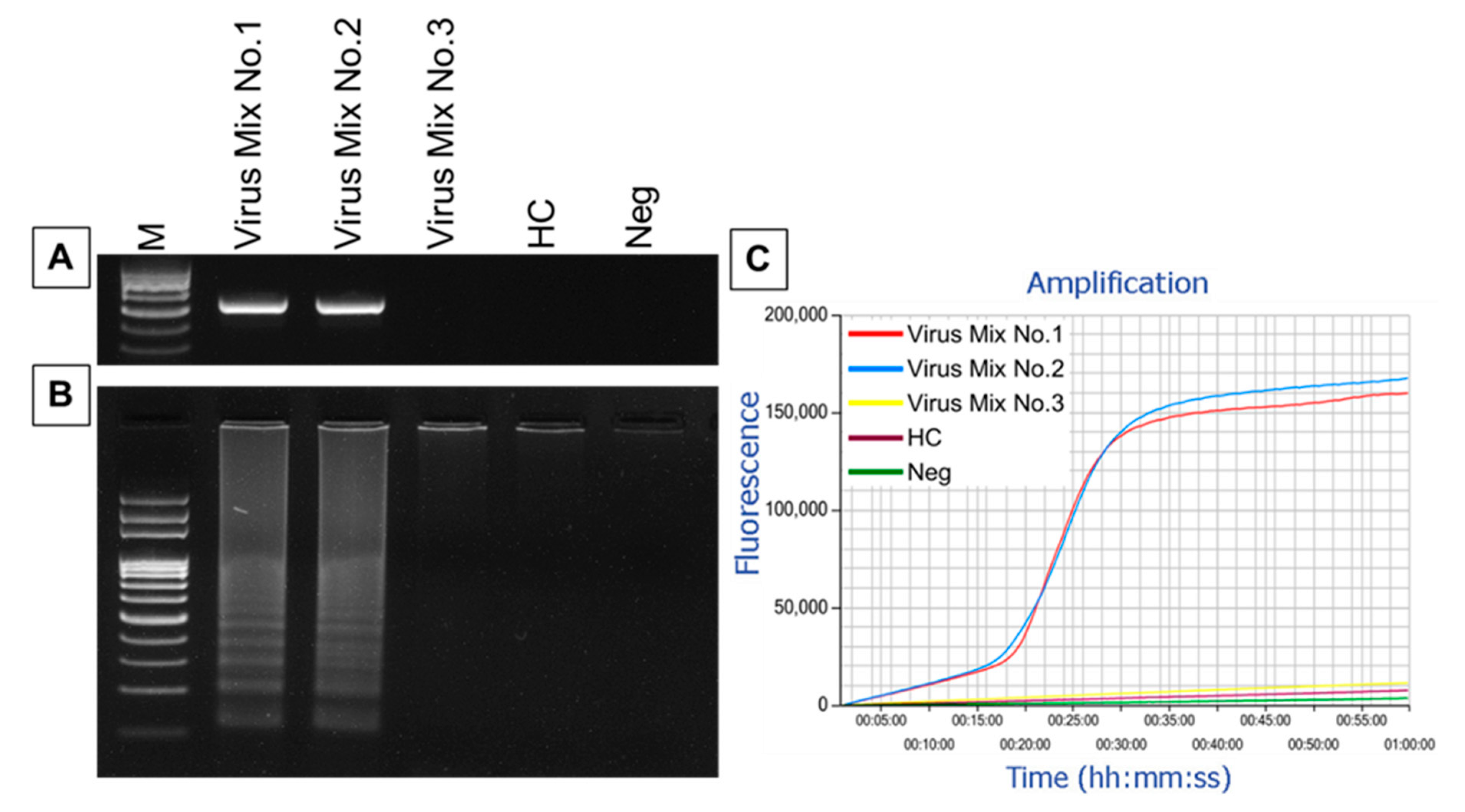
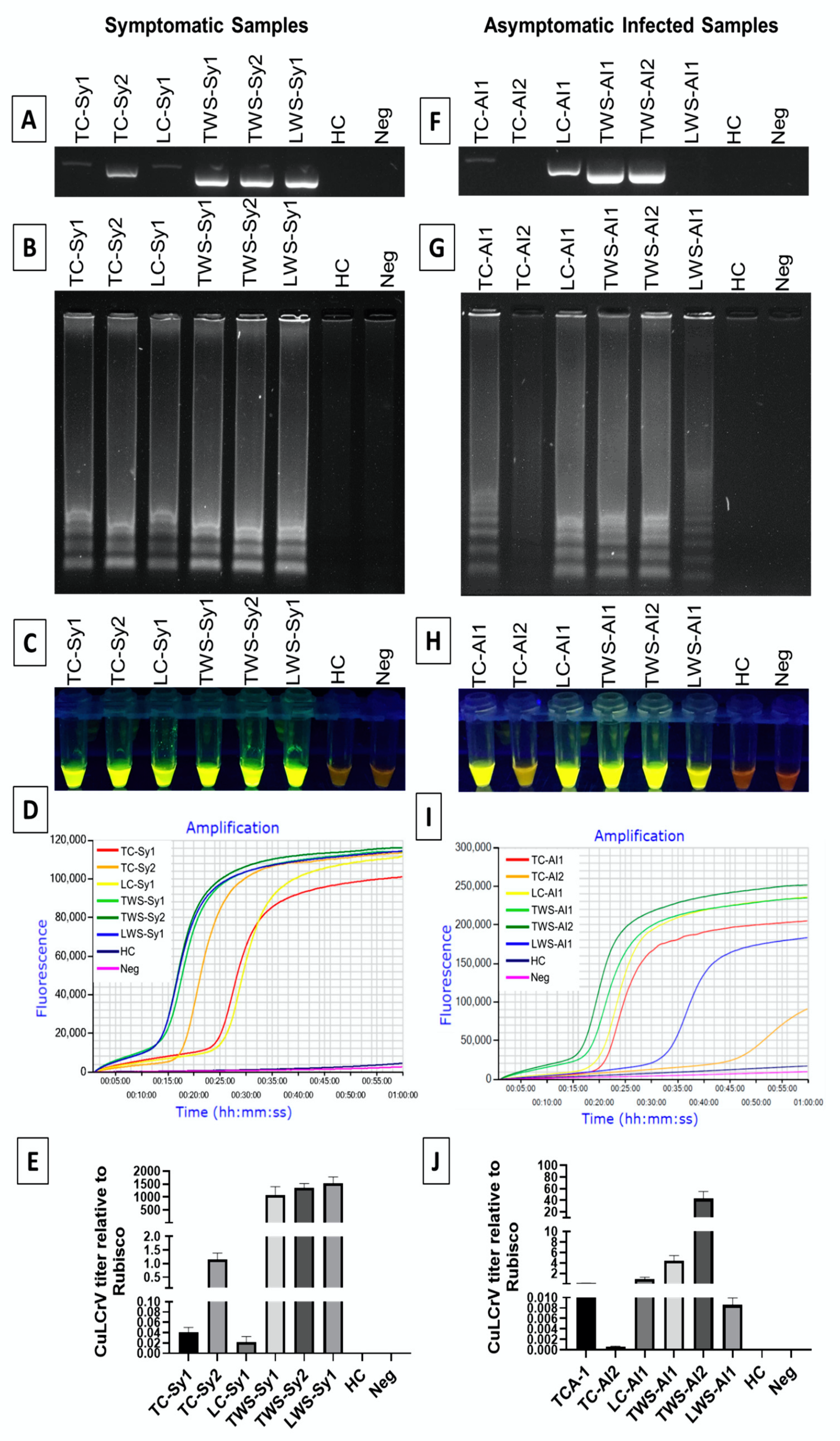
Enveloped DNA viruses can obtain their envelope by budding through the nuclear membrane or by making their way to the cytoplasm and budding into cytoplasmic vesicles or through the plasma membrane. The RT-LAMP assay is very simple and rapid and the amplification can be completed within 50 min under isothermal conditions at 63C by a set of 6 primers targeting the E gene based on the sequences analysis of the newly isolated viruses and other closely related FlavivirusThe monitoring of gene amplification can also be visualized by using SYBR green I. Only optical fluoresce microscopes can see inside a virus and then only indirectly using dye which cannot actually penetrate a virus.
Also viral particles can be easily visualized using electron microscopy and fluorescently tagged virions can be individually tracked in living cells. These virions have a sedimentation coefficient of 140165 S20w. While powerful these.
Virus-induced gene silencing VIGS is a method that takes advantage of the plant RNAi-mediated antiviral defense mechanism. Accuracy in virus quantification can be improved by using a scanning transmission electron microscopy detector STEM Hartel et al 1996 in a scanning electron microscope SEM Blancett et al 2017. However bacteriophages viruses that infect bacteria have a unique shape with a geometric head and filamentous tail fibers.
Then the mouse anti-PEDV N IgG primary antibody 1200 dilutions prepared in our. This method is entirely novel. The identical protein subunits on the virus surface can thus be labeled with an amine reactive dye and visualized through immunofluorescent microscopy.
Fluorescence microscopy can also be used to study the viability of bacterial cells. Single particles of larger viruses and helical viruses were eventually visualized by AFM and these included tobacco mosaic virus cauliflower mosaic virus Tipula iridescent virus herpes simplex virus vaccinia virus 17 36 and the largest virus of all mimivirus 30 48. TEM enables high resolution of virion.
After the virus sheds its outer coat fluorescent azide molecules bind to the ethynyls 4 offering a way to visually track individual viral genomes in the cytosol and. One common method is adding ethidium bromide a stain that inserts into the nucleic acids at non-specific locations and can be visualized when exposed to ultraviolet light. Using conventional electron microscopy the structure of the capsid surface can be visualized.
The viral DNA then incorporates ethynyl-tagged nucleosides during replication 2. Viruses replicate outside of the cell. Other stains that are safer than ethidium bromide a potential carcinogen.
The Baltimore Classification system describes viruses according to which characteristic. Acapsid shape bhost-cell susceptibility. Its as if the.
Using fluorescence microscopy to study bacterial cell viability. The virus particles contain a single species of ssRNA. For WNV virus genomics played a central role in uncovering how the virus emerged and became endemic in a new region after its introduction in the US eg 2127 and more detailed studies may identify transmission networks and source areas of potential reemergence to be targeted during future interventions 2829.
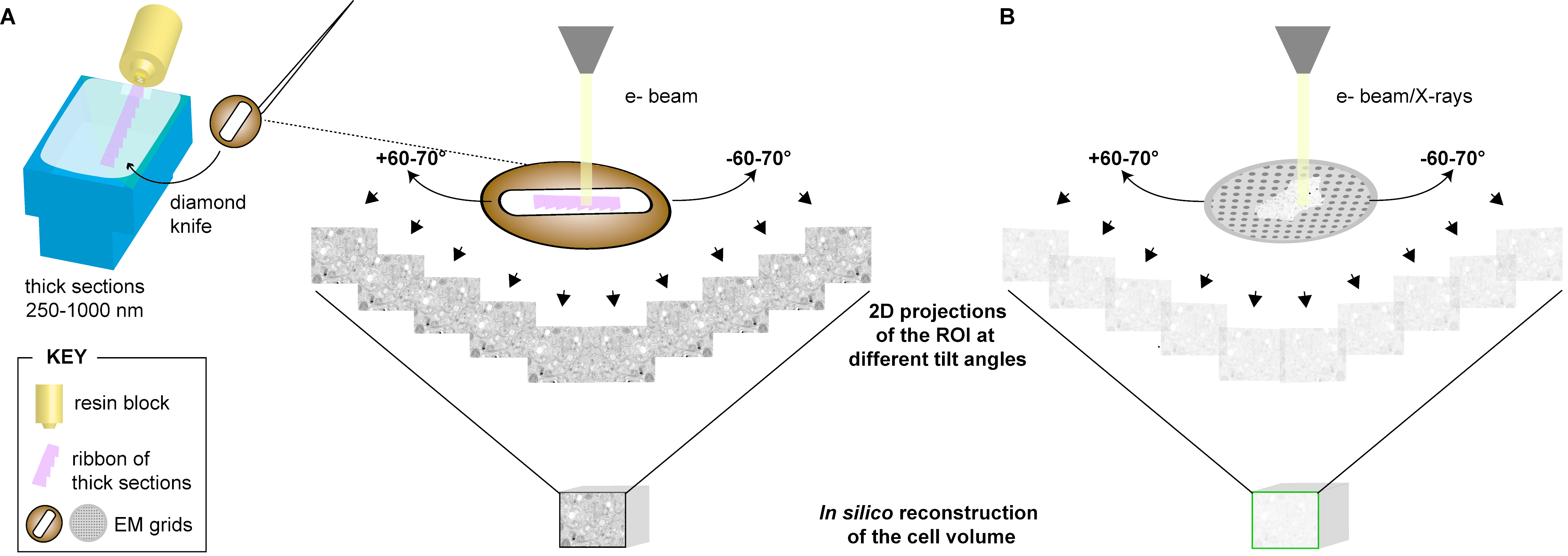
The surface structure of virions can be observed by both scanning and transmission electron microscopy whereas the internal structures of the virus can only be observed in images from a transmission electron microscope Figure 124. Sigma USA to promote the adhesion of virus particles to glass surfaces. Can easily make a virus model from paper with proteins printed on the surface.
An alternative label is an enzyme that can be used to generate many molecules of detectable product by catalyzing the conversion of substrate molecules. Here we present a simple method of labeling of dengue virus with Alexa Fluor succinimidyl ester dye dissolved directly in a sodium bicarbonate buffer that yielded highly viable virus after labeling. Name of virus is on virus tape and protein.
We show that the use of different imaging wavelengths allows the visualization of a multitude of virus particles. Using a previously described method for metagenomics sequencing 217 only a small percentage. Viruses can be visualized using which method.
Viruses are estimated to outnumber their cellular hosts in most environments 12 indicating that animals regularly encounter and interact with virusesThe ubiquity of viruses is demonstrated by the widespread success of virus discoveries made through metagenomic sequencing of animal feces and tissues 35While surveys of virus prevalence.
Ijms Free Full Text Development Of Loop Mediated Isothermal Amplification Assay For Rapid Detection Of Cucurbit Leaf Crumple Virus Html
Ijms Free Full Text Development Of Loop Mediated Isothermal Amplification Assay For Rapid Detection Of Cucurbit Leaf Crumple Virus Html

Optimizing The Synthesis And Purification Of Ms2 Virus Like Particles Scientific Reports
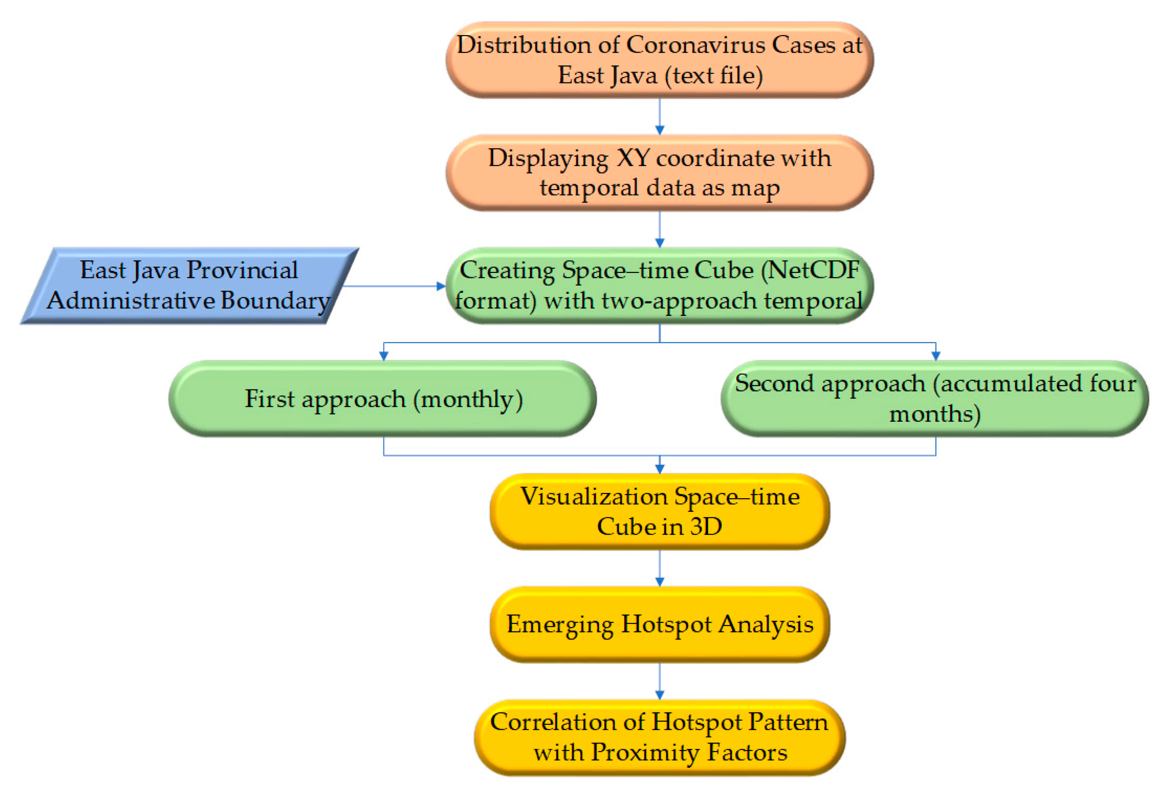
Ijgi Free Full Text Spatiotemporal Analysis Of Covid 19 Spread With Emerging Hotspot Analysis And Space Time Cube Models In East Java Indonesia Html
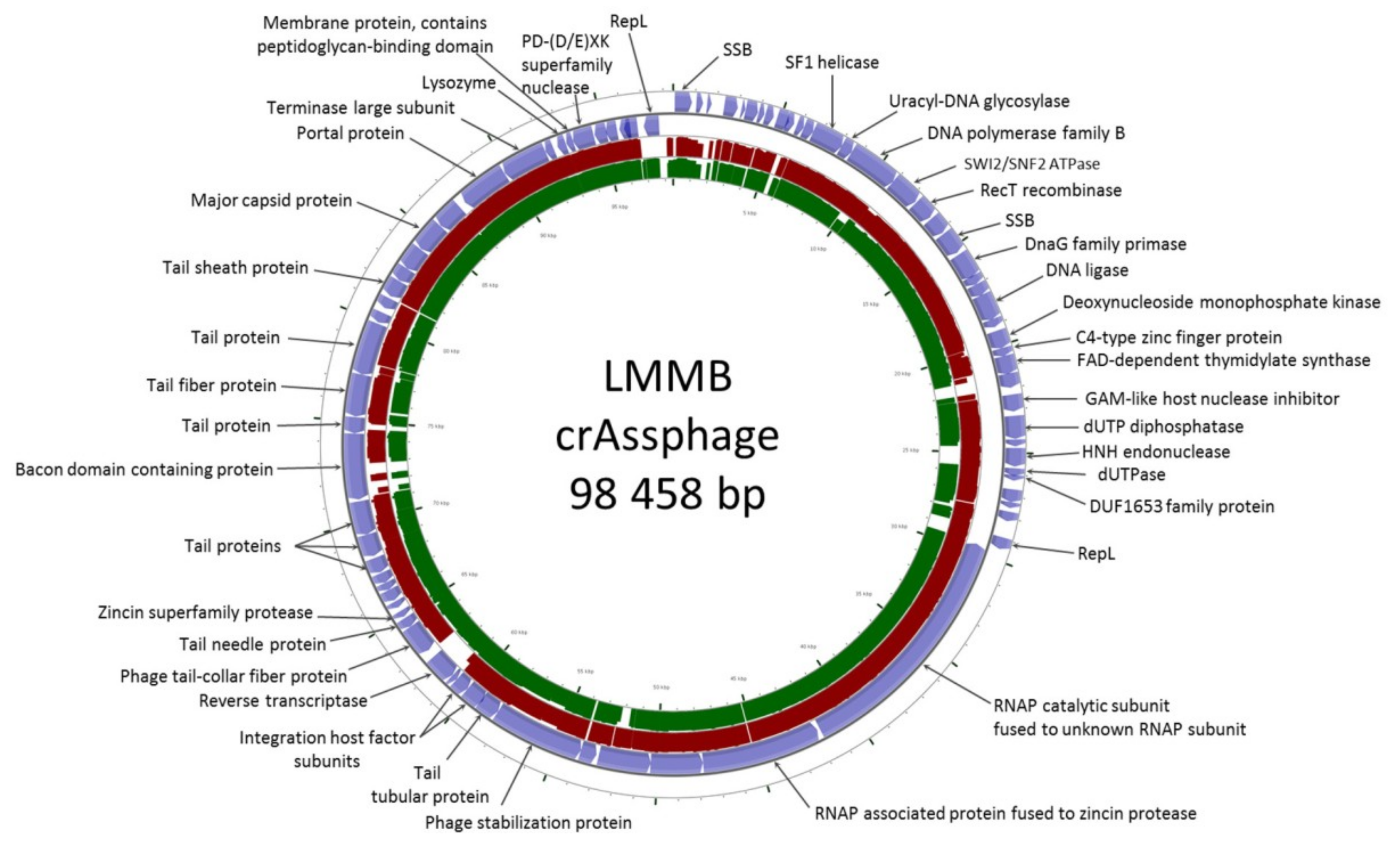
Viruses Free Full Text First Crass Like Phage Genome Encoding The Diversity Generating Retroelement Dgr Html
Ijerph Free Full Text Wastewater Based Epidemiology As An Early Warning System For The Spreading Of Sars Cov 2 And Its Mutations In The Population Html
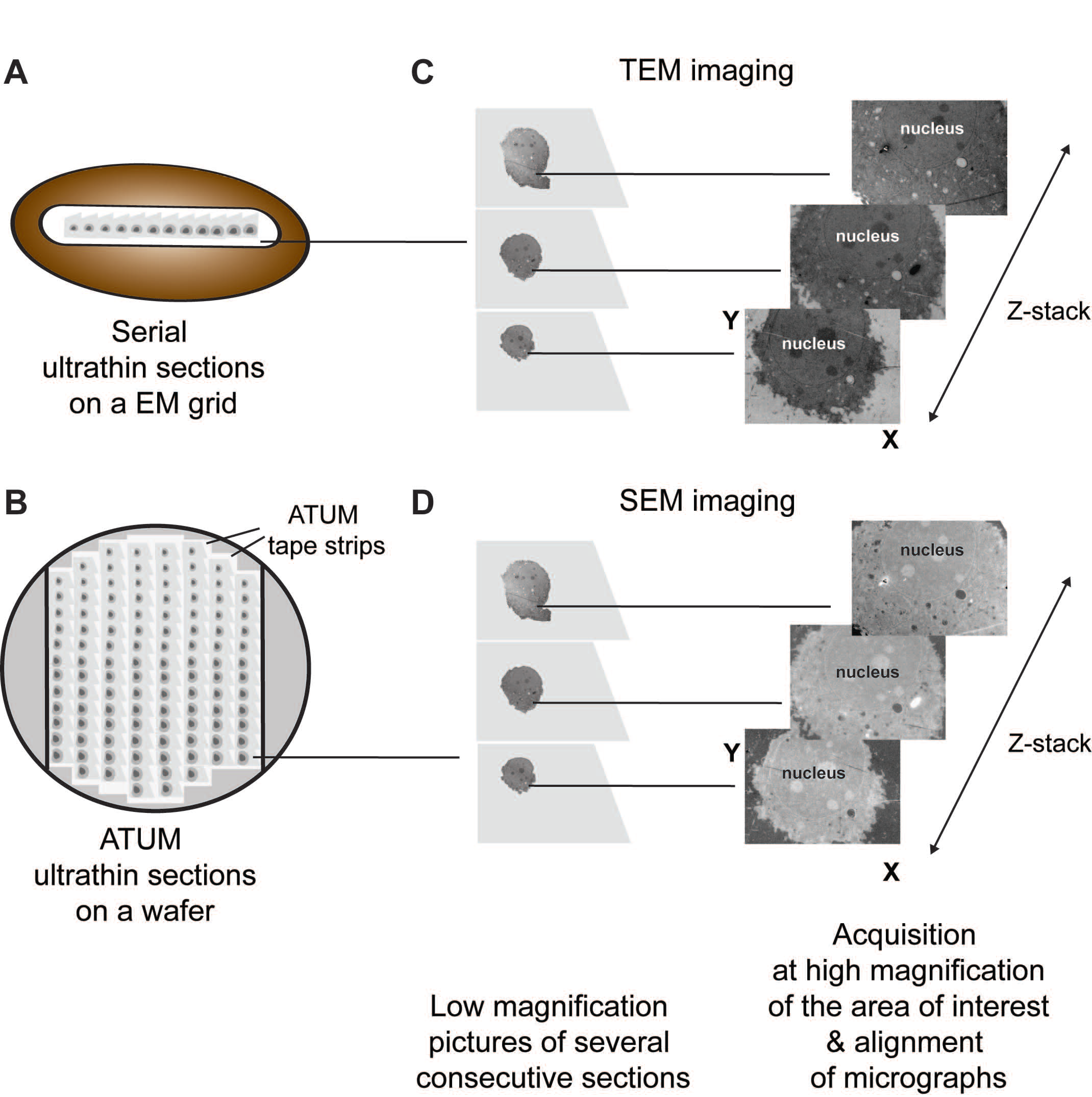
Viruses Free Full Text Viral Infection At High Magnification 3d Electron Microscopy Methods To Analyze The Architecture Of Infected Cells Html
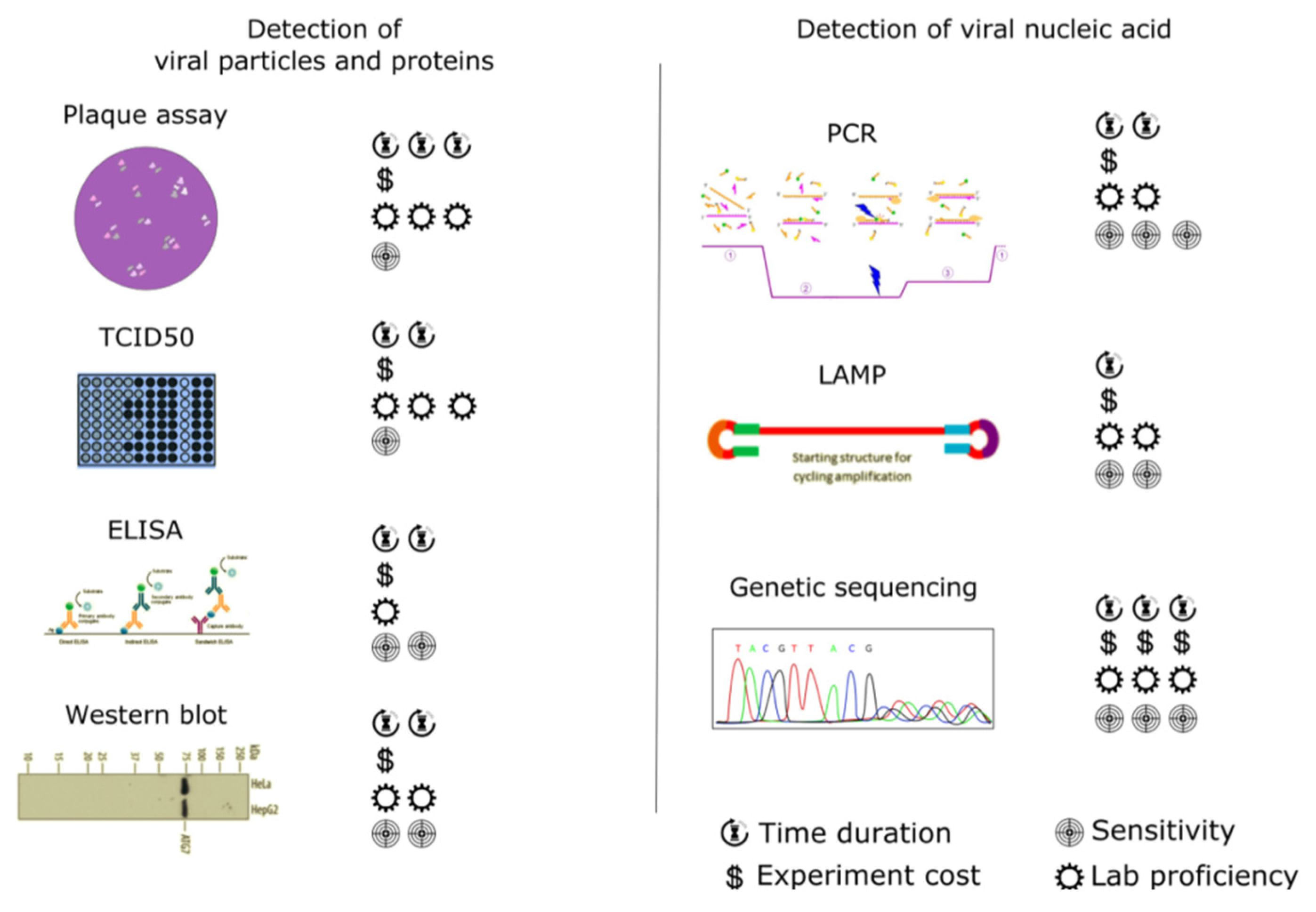
Detection Of Rna Viruses From Influenza And Hiv To Ebola And Sars Cov 2 A Review Analytical Methods Rsc Publishing Doi 10 1039 D0ay01886d
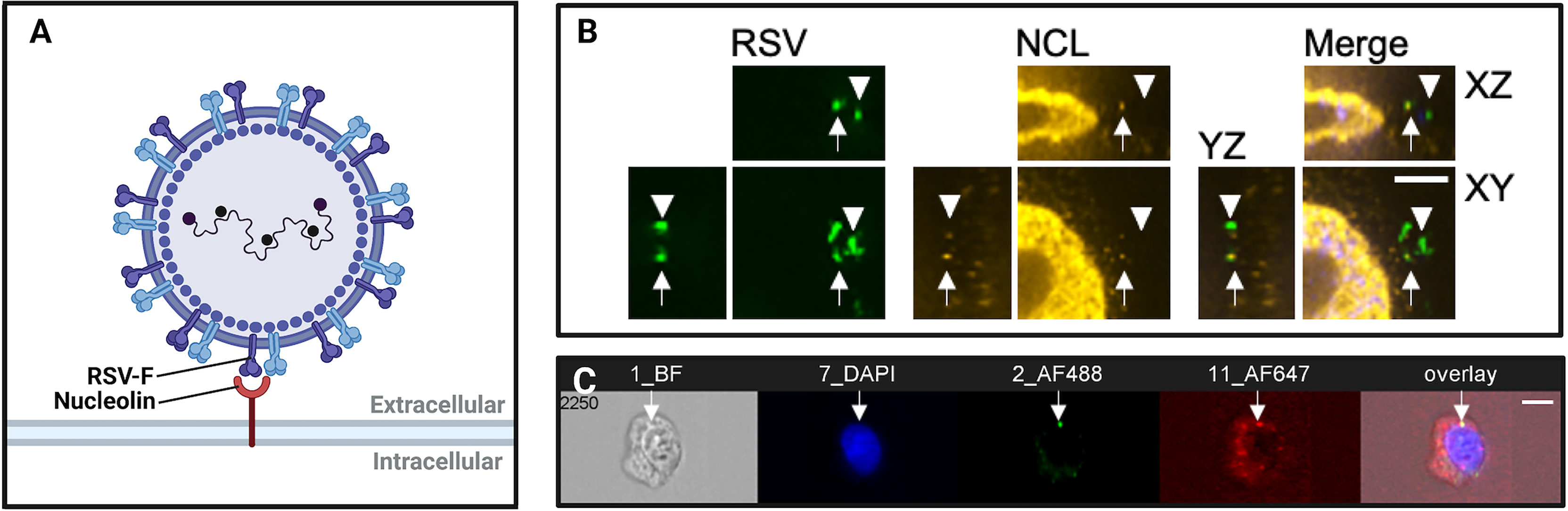
Frontiers Imaging Flow Cytometry And Confocal Immunofluorescence Microscopy Of Virus Host Cell Interactions Cellular And Infection Microbiology
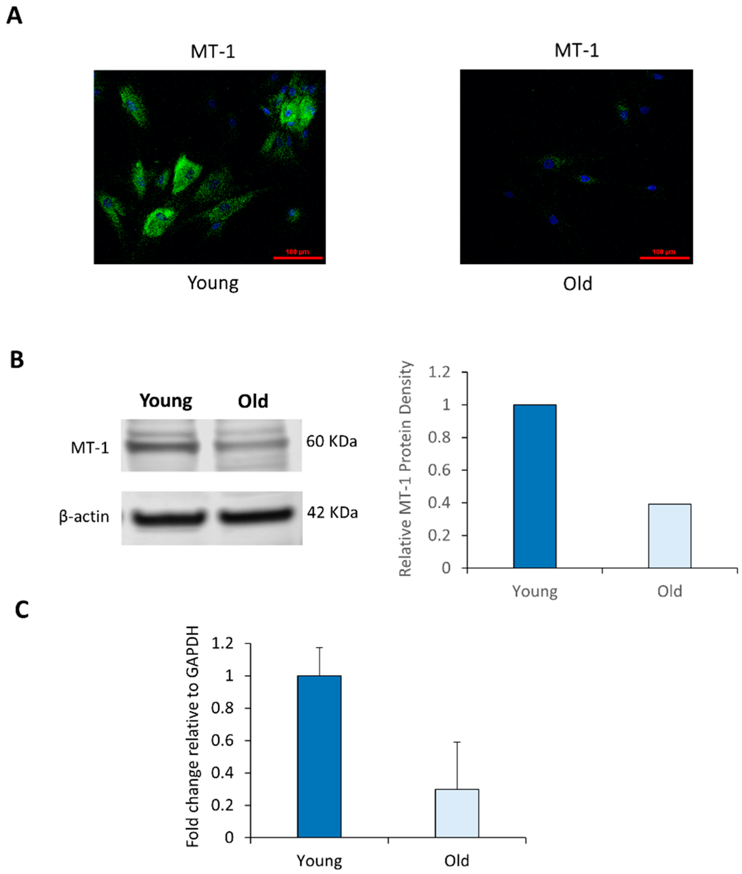
Ijms Free Full Text Age Associated Decrease Of Mt 1 Melatonin Receptor In Human Dermal Skin Fibroblasts Impairs Protection Against Uv Induced Dna Damage Html

Microorganisms Free Full Text Phylum Gemmatimonadota And Its Role In The Environment Html
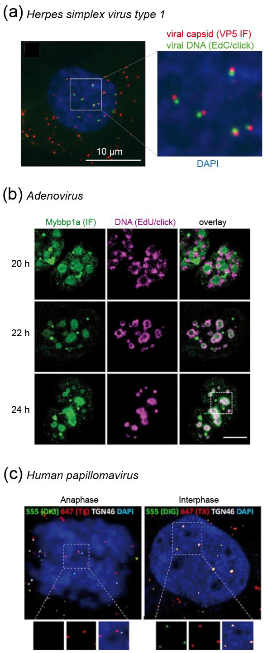
Molecules Free Full Text A Spotlight On Viruses Application Of Click Chemistry To Visualize Virus Cell Interactions Html

Ijgi Free Full Text Spatiotemporal Analysis Of Covid 19 Spread With Emerging Hotspot Analysis And Space Time Cube Models In East Java Indonesia Html

Investigating The Concept And Origin Of Viruses Trends In Microbiology

Ijms Free Full Text Development Of Loop Mediated Isothermal Amplification Assay For Rapid Detection Of Cucurbit Leaf Crumple Virus Html
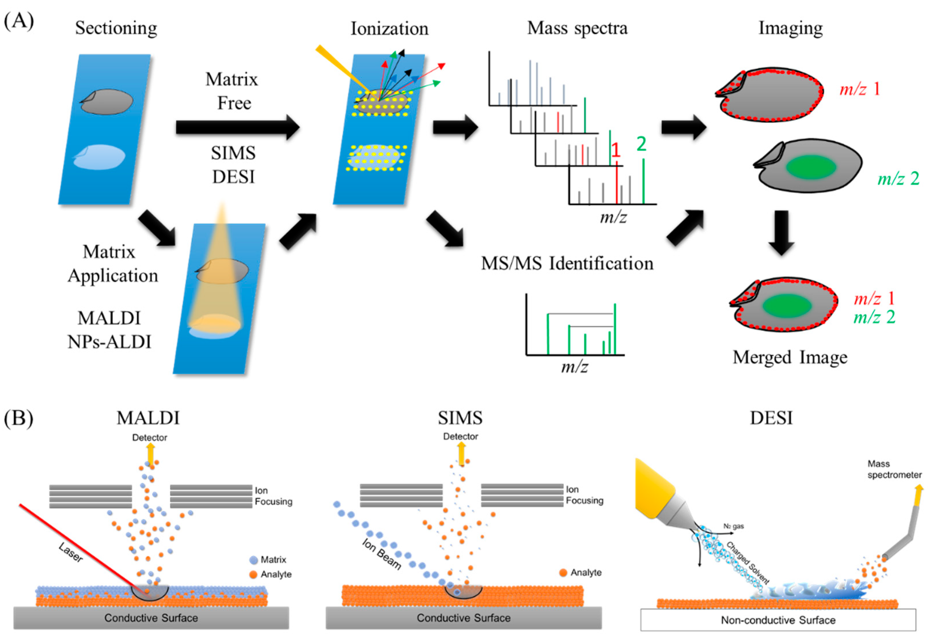
Foods Free Full Text Application Of Mass Spectrometry Imaging For Visualizing Food Components Html
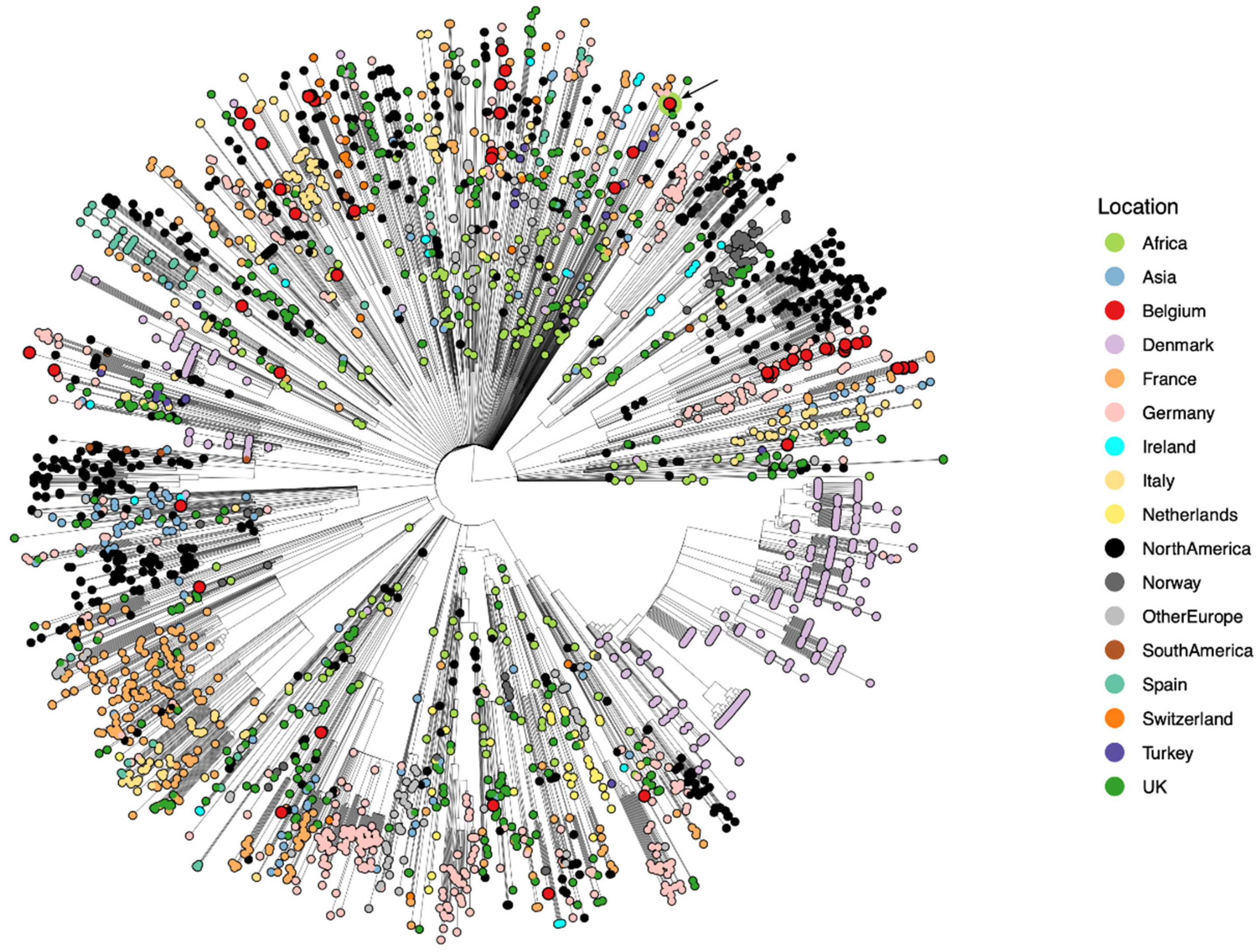
Viruses Free Full Text Variant Analysis Of Sars Cov 2 Genomes From Belgian Military Personnel Engaged In Overseas Missions And Operations Html
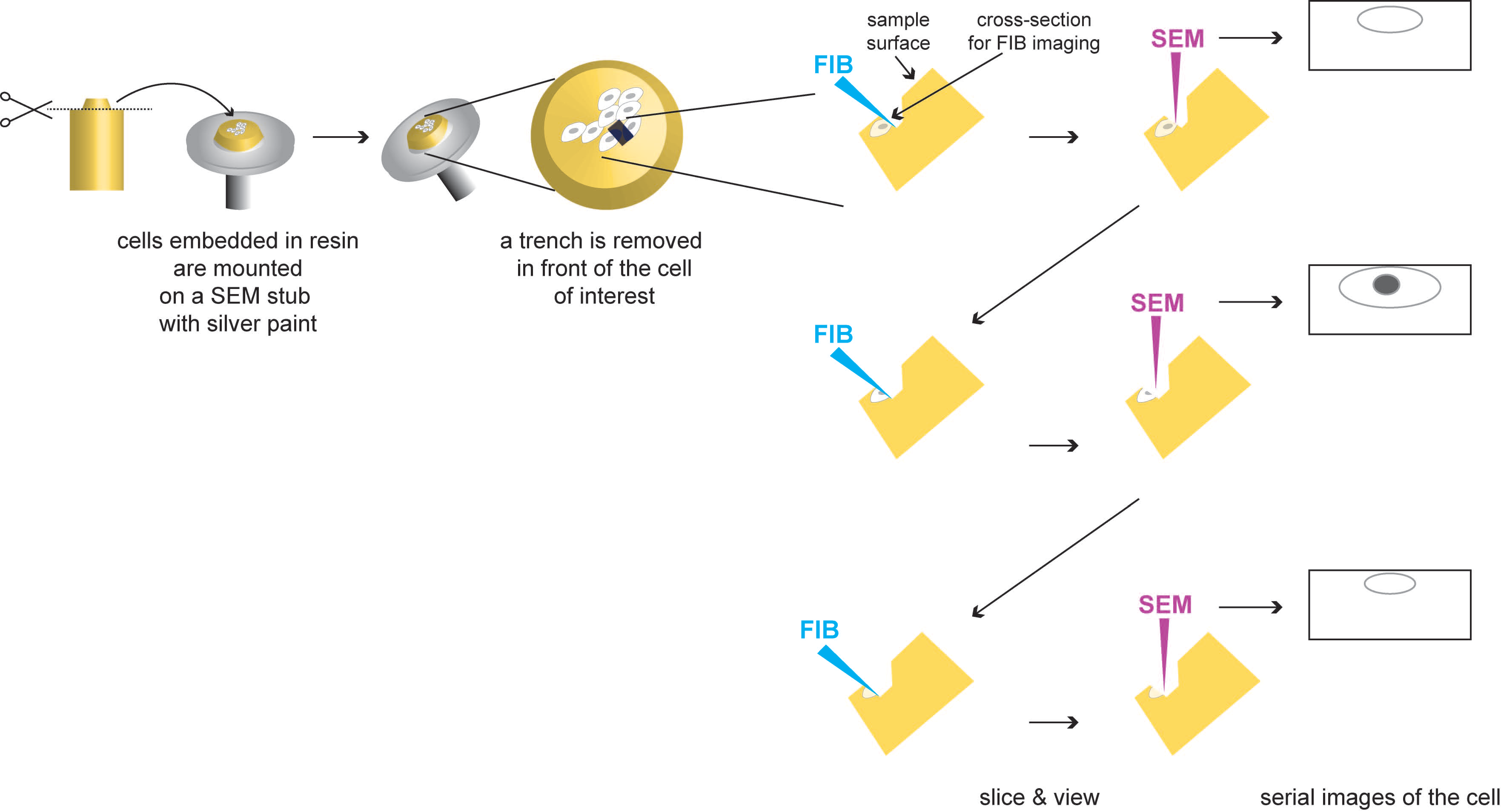
Viruses Free Full Text Viral Infection At High Magnification 3d Electron Microscopy Methods To Analyze The Architecture Of Infected Cells Html

Enzymatic Assays To Explore Viral Mrna Capping Machinery Kasprzyk 2021 Chembiochem Wiley Online Library


Comments
Post a Comment